|
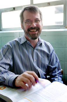
Dr. D. Brian Thompson |
||
Associate Professor of Physics |
|||
B.S., 1987, Kentucky Wesleyan College
|
|||
|
Office: 219 Floyd Science Building
|
|||
|
|
Dr. D. Brian Thompson |
||
Associate Professor of Physics |
|||
B.S., 1987, Kentucky Wesleyan College
|
|||
|
Office: 219 Floyd Science Building
|
|||
Brian joined the UNA faculty in 1999 after working at synchrotron radiation laboratories in the United States, England, and Italy. At UNA, he has built an optical tweezers used to trap and manipulate micron-sized particles. To see recent applications of the optical tweezers, choose from the movie list below.
Moving a particle .wmv (2.7 MB) .mpg (3.7 MB)
Manually positioning a 2 micrometer latex microsphere along a square path. The out-of-focus microspheres not moving in the upper-left of the image are stuck by surface tension to the coverslip of the microscope slide.
Rotating a particle .wmv (6.5 MB) .mpg (9.3 MB)
Circularly-polarized light rotates a birefringent calcite particle of length about 1 micrometer. The trapped particle is located at the center of the image. Other calcite pieces can be seen floating by.
Trapping a particle .wmv (6.6 MB) .mpg (9.3 MB)
Moving the microscope slide in order to trap a calcite particle. The optical trap is located at the center of the image. Out-of-focus blobs that don’t move are calcite particles stuck to the coverslip. When the calcite particle is tracked down and trapped, it immediately begins rotating.
Trapping two particles .wmv (4.0 MB) .mpg (5.9 MB)
The incident laser beam is split to form two traps that can be moved independently or together. The image shows two 2-micrometer latex microspheres trapped at the center of the image. Initially, the two particles are moved about together, then they are moved about separately.
Two particles dancing .wmv (10.4 MB) .mpg (15.4 MB)
Another demonstration that two particles can be moved independently.
Computer-controlled positioning of a particle .wmv (2.2 MB) .mpg (3.1 MB)
A computer is used to position a 5 micrometer latex microsphere from a central position, along a square path, and back to the central postion. The out-of-focus microsphere not moving in the upper-left of the image is stuck on the coverslip of the microscope slide. The computer-controlled actuators can position the optical trap with about a 10 nm step resolution.
Live animals, spinning bacterium .wmv (6.1 MB) .mpg (8.9 MB)
A long, flexible bacterium is tracked down and trapped. The optical trap is located at the center of the image. The bacterium spins around as the trap holds it at one end. An infrared laser was used to trap the bacterium, so that it was not harmed.
Live animals, rigid bacterium .wmv (2.7 MB) .mpg (3.8 MB)
A long, rigid bacterium is tracked down and trapped. The optical trap is located at the center of the image. The trap is used to drag the bacterium about.
Live animals, trapped protist .wmv (6.0 MB) .mpg (8.8 MB)
A protist with a single(?) flagellum thrashes about in the optical trap, eventually escaping, only to be recaptured. The optical trap is located at the center of the image. Note the two small bacteria, stacked one on top of the other, hitching a ride on the protist.
Coupling fluorescent microspheres .wmv (2.2 MB) .mpg (13.3 MB)
Two 6 um microspheres, ring-stained with a fluorescence dye, held in separate traps. Less than 0.1 mW of power from the 488 nm blue beam of an argon-ion laser is focused in a trapping configuration, located one particle diameter to the right of the rightmost sphere. This beam (not visible in the picture) excites the dye, causing the spheres to fluoresce. The resulting fluorescence spectra exhibit resonance lines that split as one sphere is brought close to the other.
Another coupling of fluorescent microspheres .wmv (1.8 MB) .mpg (12.1 MB)
Another pair of 6 um fluorescent microspheres held in separate traps, along with an argon ion beam, as detailed above. As the leftward sphere is brought toward the stationary rightward sphere and the argon-ion beam, of course its fluorescence increases. But watch for the significant decrease in the leftward sphere's fluorescence when the rightward sphere is allowed to escape from its trap.
| Physics and Earth Science Homepage | Faculty | Requirements for Degrees |
E-mail comments to Dr. Mark Puckett
Last update August 1, 2006
Return to UNA Homepage